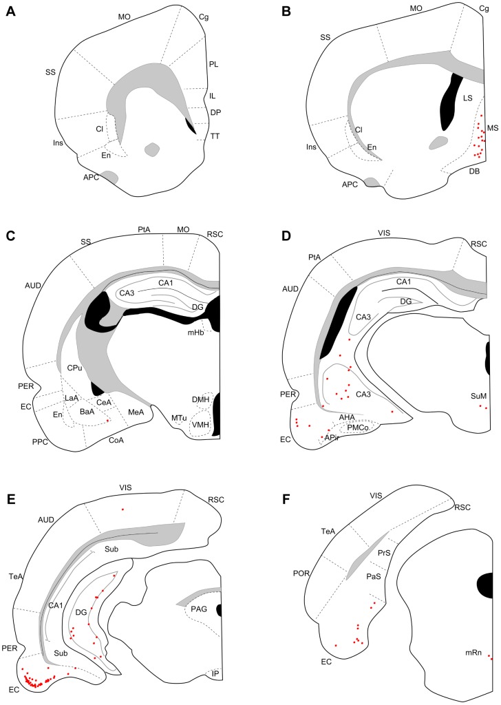
Figure 3. Distribution of retrogradely labeled neurons three days after virus injections into ventral DG.
Series of coronal sections (organized from rostral to caudal) for a rat surviving three days after ventral DG injection. AHA, amygdalohippocampal area; PMCo, posteromedial cortical nucleus of the amygdala. Remainder of abbreviations the same as in Figure 2.

