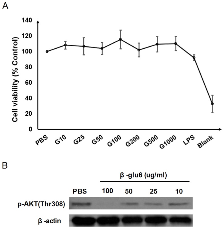
Figure 1. β-glu6 (G) suppresses AKT phosphorylation and does not affect peritoneal macrophage viability.

The peritoneal macrophages were treated with 10 μg/mL, 25 μg/mL, 50 μg/mL, 100 μg/mL, 200 μg/mL, 500 μg/mL and 1000 μg/mL of β-glu6 (G) or 1 μg/mL of LPS, and the viability of the macrophages was examined by the MTT assay, as described in the Materials and Methods section. Data are expressed as the mean ± SD of three independent experiments (A). Macrophages were treated with 10 μg/mL, 25 μg/mL, 50 μg/mL, and 100 μg/mL of β-glu6 (G) for 2 h, and the level of phosphorylated AKT (Thr308) was detected by western blot assay. Beta-actin was used as a loading control. Data shown represent one of three independent experiments (B).
