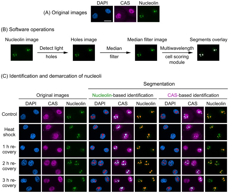
Figure 1. Nucleolin demarcates nucleoli upon heat shock and during the recovery from heat stress.
(A) Original images for unstressed cells. (B) Software operations applied to detect nucleoli. The original image of the marker that demarcates the nucleolus (i.e. nucleolin) is subjected to a set of operations to define nucleoli and create nucleolar segments. In brief, the “detect light holes” filter identifies nucleoli based on the difference in fluorescence intensities between nucleoli and the surrounding nucleoplasm; this generates a “holes image”. The holes image is then passed through the “median filter” to reduce noise, generating the “median filter image”. The multiwavelength cell scoring module uses the median filter image as a template to produce segments that colocalize with nucleoli. The module accurately demarcates the boundaries of nucleoli on the basis of size constraints and intensity above local background [21]. (C) Demarcation of nucleoli in heat-stressed cells. HeLa cells were exposed to severe heat shock (1hour at 45.5°C), fixed immediately or after recovery at 37°C for the times indicated. Control samples were not heat-stressed. Fixed cells were processed for immunofluorescence staining with antibodies against nucleolin and CAS; nuclei were stained with DAPI. Confocal images were employed to identify and demarcate nucleoli with MetaXpress image analysis software. Segmentation and segments overlay panels depict the identification of nucleoli. Note that nucleolin delimited the nucleoli successfully under all conditions (Nucleolin-based identification). By contrast, CAS was not a suitable marker for heat-shocked cells until 3 hours recovery. Size bar is 20 µm.

