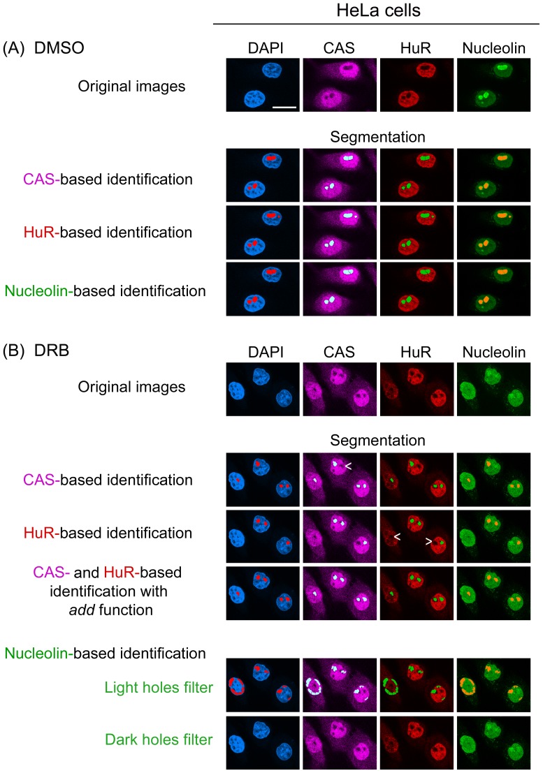
Figure 5. CAS and HuR, but not nucleolin, delimit nucleoli in DRB-treated HeLa cells.
Cells were incubated with (A) DMSO or (B) DRB essentially as described [21] and processed as in Fig. 2. Individually, the marker proteins CAS and HuR detected nucleoli upon DRB incubation, although some nucleoli were missed (indicated by arrow heads). The identification of nucleoli was improved by combining the information from CAS and HuR images with the add function [21]. Nucleolin was redistributed by DRB throughout the nucleoplasm. Based on the nucleolin image, neither the “detect light holes” nor “detect dark holes” filter could identify nucleoli. Size bar is 20 µm.

