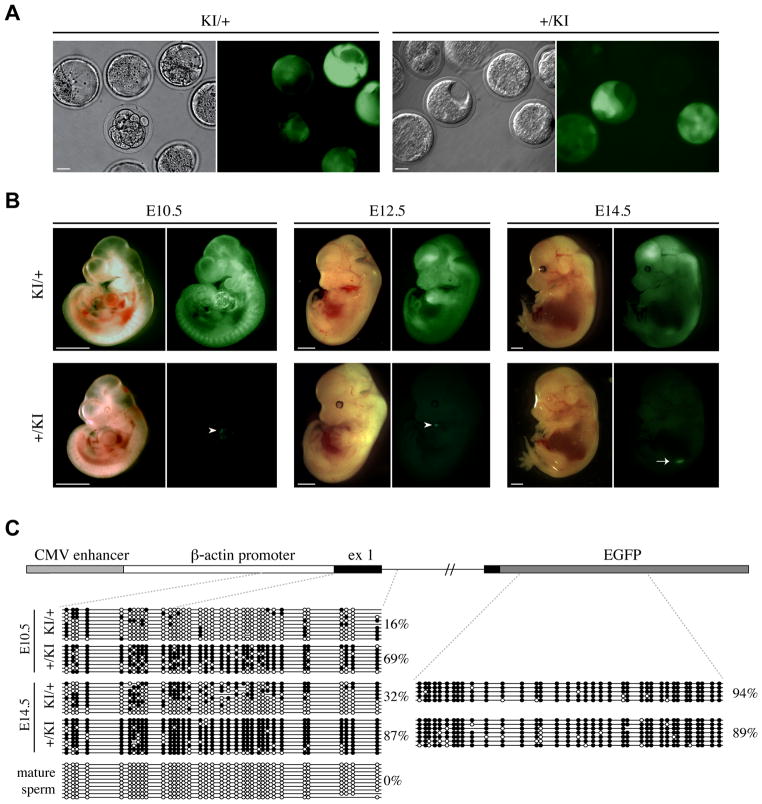
Fig. 2. Relationship between imprinted GFP expression and DNA methylation at the CAG promoter in post-implantation embryos.
(A) E3.5 maternal (KI/+) and paternal (+/KI) heterozygous embryos were collected from crosses between wild-type C57BL/6J and Tel7KI (KI for short) heterozygous mice. Pre-implantation embryos were observed by microscopy under bright field (left) or GFP fluorescence (right). Scale bar: 20 μm. (B) Imprinted expression of Tel7KI in post-implantation embryo. E10.5, E12.5, and E14.5 embryos carrying a maternal (KI/+) or a paternal (+/KI) Tel7KI were visualized under bright field (left) or GFP fluorescence (right). Occasional GFP fluorescence is observed in hearts of +/KI embryos (white arrowheads), and GFP expression from the gonads of +/KI embryos can be seen through the body wall after E13.5 (white arrow). Scale bar: 1 mm. (C) Structure of the CAG-EGFP reporter of Tel7KI (above) showing the CMV enhancer, the chicken β-actin promoter, and a 5′ intron from the chicken β-globin gene, fused to EGFP. Sequencing of sodium bisulfite-modified genomic DNA (below) purified from paternal (+/KI) and maternal (KI/+) transmission embryos and mature sperm from a +/KI adult male. Circles represent methylated (filled) or unmethylated (open) CpGs; absent circles indicate ambiguous sites.

