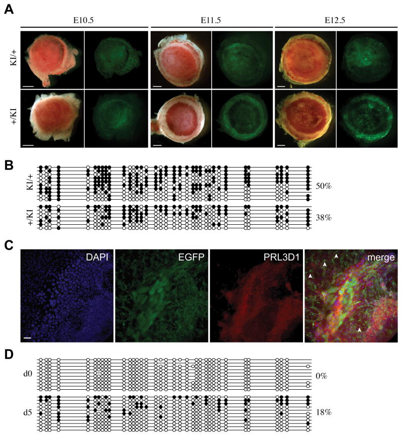
Fig. 5. Lack of imprinted GFP expression and DNA methylation at Tel7KI in the placenta and in cultured trophoblast giant cells.
(A) Placentae carrying maternal (KI/+) or paternal (+/KI) alleles of Tel7KI visualized under bright field (left) and GFP fluorescence (right) at E10.5, E11.5, and E12.5. Scale bars: 1 mm. (B) Promoter DNA methylation analysis by bisulfite sequencing at Tel7KI in whole E14.5 placentae carrying a maternal (KI/+) or paternal (+/KI) Tel7KI. Filled circles represent methylated CpGs, open circles represent unmethylated CpGs and absent circles are CpGs for which data is unavailable. (C) Immunohistochemical detection of GFP in giant cells. Trophoblast giant cells (TGCs) grown from EPCs in culture for 5 days were stained with antibodies against GFP and the TGC marker placental lactogen 1 (PRL3D1). Scale bar: 100 μm. (D) DNA methylation analysis of paternal transmission Tel7KI E8.5 EPCs both uncultured (d0) and cultured in vitro for 5 days (d5). Sodium bisulfite-modified genomic DNA was analyzed for DNA methylation patterns at the CAG promoter of the paternal Tel7KI allele.

