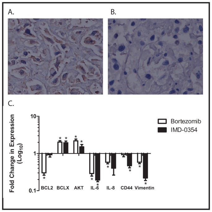
Figure 5. A) Immunohistochemical analysis of sections of xenograft from control mice showing expression of NF-κB p65 (brown staining).
(B) Immunohistochemical analysis of sections of xenograft from mice treated with bortezomib, also stained with antibody for the p65 subunit of NF-κB. No staining is evident in these sections. Images are at 200x magnification. C) RNA was isolated from xenografts growing in untreated, control mice, and in mice treated with either bortezomib or IMD-0354. After reverse transcription, quantitative RT-PCR was performed to measure expression of the indicated NF-κB target genes. Data are presented as fold change between mice treated with the indicated agent and control. Experiments were performed in triplicate, and error bars show standard error of the mean. *=achieves statistical significance compared to control (p<0.05) by two-tailed unpaired Student t test.

