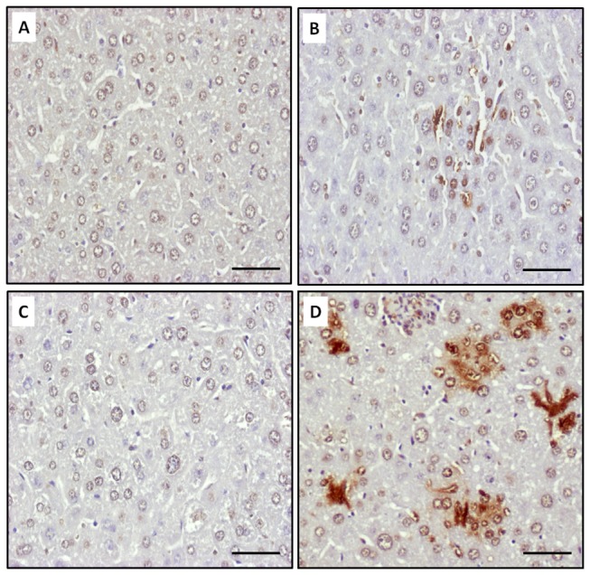
Figure 7. K. pneumoniae-induced hepatic apoptosis.

Liver sections of PBS-control naïve mice (A), K. pneumoniae-infected naïve mice (B), PBS-control diabetic mice (C), and K. pneumoniae-infected diabetic mice (D) were subjected to TUNEL analysis. The nuclei of apoptotic cells have been stained brown with the TUNEL method. Scale bar represents a distance of 50 μm.
