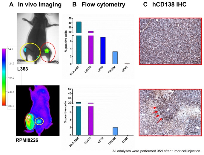
Figure 2. A. Detection of human L363 and RPMI8226 cells in immunodeficient mice using hCD138 antibody labelled with Alexa750.
20 mice per cell line were engrafted with L363 or RPMI8226 cells via different application routes. Images were taken once weekly for five weeks after implantation of the respective MM cell line with the Kodak Image Station in vivoFX. In addition to the determination of tumor load via CCD camera, animals were x-rayed and the two pictures merged for optimal localization of the fluorescent region. No x-ray was performed for the animal bearing RPMI8226 in this figure. Intraosseal tumor growth was clearly detectable by the in vivo imaging (IVI) system (red circles). Additionally, BM metastases became apparent within the adjacent tibia and lumbar spine (yellow circles). B. Detection of human L363 and RPMI8226 cells in immunodeficient mice using flow cytometry. IVI data for L363 and RPMI8226 were confirmed by flow cytometry 35 days after tumor cell injection showing 42% and 22% human HLA-ABC and CD138 positive cells in L363 and 21% and 20% for RPMI8226, respectively. Analyses were performed on metastatic BM lesions. C. Detection of human L363 and RPMI8226 cells in immunodeficient mice using immunohistochemistry. IVI data for L363 and RPMI 8226 cells were confirmed by immunohistochemistry specific for human CD138+ cells (CD138+-infiltrates, red arrows) Analyses were performed on metastatic BM lesions.

