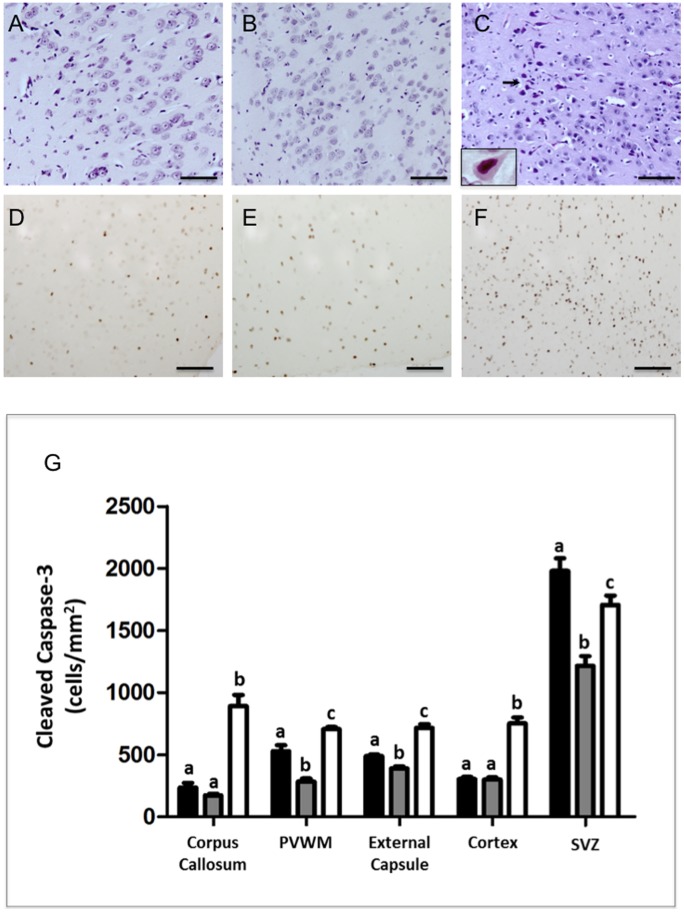
Figure 2. Hematoxylin & Eosin (A–C).
Few degenerating cells were seen in the cortical gray matter of full-term (A) and preterm (B) control lambs. UCO lambs showed extensive neuronal injury displaying feature of apoptosis seen by H&E staining as scattered dark, shrunken cells with pyknotic or small, densely staining nuclei and eosinophilic cytoplasm (black arrows C). The insert in panel C is a high power micrograph showing a neuron exhibiting ischemic morphology (eosinophilia, and nuclear pyknosis). Panels D–F show activated Caspase-3 immunoreativity in the cortex of a full term (D), preterm (E) and UCO (F) lambs. Note the increased immunoreactivity of activated Caspase-3 in the cortex of UCO lambs compared with both control groups. Scale bars –50 µm. Panel G: Quantitative results show cleaved caspase-3 cell counts in the corpus callosum, periventricular white matter (PVWM), external capsule, cortex and subventricular zone (SVZ) for full-term control (n = 5), preterm control (n = 5) and UCO lambs (n = 6) (G). Data are expressed as mean ± SEM. p<0.05. White, gray and black bars represent full term, pre term and UCO lambs respectively.

