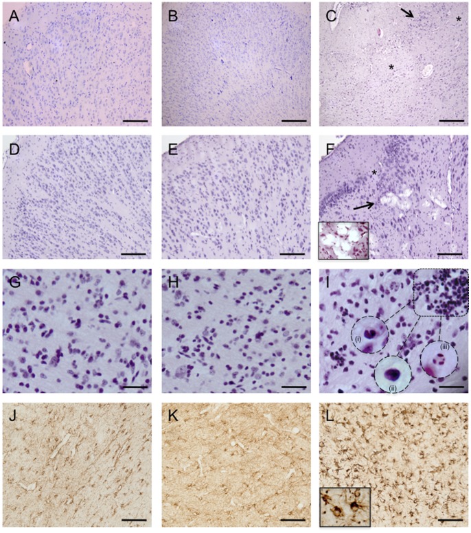
Figure 3. Hematoxylin & Eosin staining of full term control (A, D & G), preterm control (B, E & H), and UCO lambs (C, F &I).
Normal cytoarchitecture of the cerebral cortex was seen in both full-term (A) and preterm brains (B). In UCO brains (C) subtle alterations in the cellular composition and spatial arrangement of neurons was seen throughout the cortical gray matter. Extensive neuronal injury (arrows) in the cortical gray matter of UCO lambs as well as areas devoid of neurons (asterisks). D & E show normal pathology in the cortex of full term and preterm control lambs respectively. In UCO lambs (F) some other cellular degenerative changes were observed, such as vacuolation of brain parenchyma (arrow). The insert in panel F shows a high power view of the vacuolar degeneration, histologic features consistent with hypoxic/ischemic changes. Inflammatory cell infiltration was seen in the periventricular white matter of UCO lambs (I), as marked by the box, which showed the morphological appearance of eosinophils (i), lymphocytes (ii) and neutrophils (iii) (inserts in I). These were not seen in full term (G) or preterm control lambs (H). Panels J, K & L are representative images of glial fibrillary acidic protein (GFAP) staining in the periventricular white matter of full term control, preterm control and UCO lambs respectively. UCO lambs displayed reactive astrocytosis. Note the dense staining of the enlarged cell bodies and the highlighted cell processes shown in the high power insert in panel L. Scale bars: A–F = 100 µm, G–L = 50 µm.

