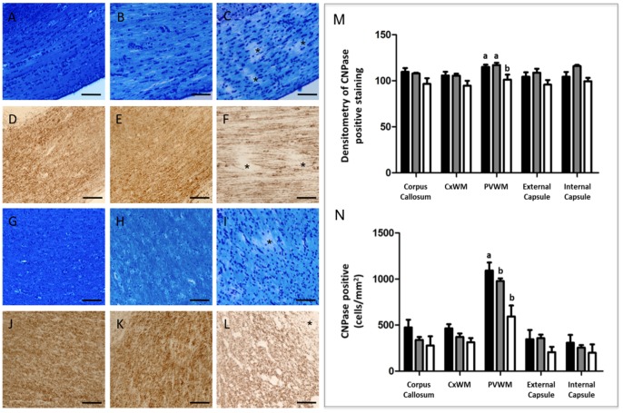
Figure 4. Luxol fast blue staining on brain sections of full term control (A), preterm control (B) and UCO lamb (C) in the corpus callosum.
Myelin irregularities (disruption) seen as patchiness (asterisks in C) were detected only in UCO lambs. Panels D–F show CNPase immunohistochemistry in the corpus callosum of a full term (D), preterm (E) and UCO lamb, confirmed myelin disruption seen as patchy staining (asterisks). Myelin disruption was also seen in the periventicular white matter of UCO lambs both with luxol fast blue (I) and CNPase (L) (asterisks). Myelination in full term (G & J) and preterm (H & K) appeared to be intact with both stains. Scale bars = 50 µm. Quantitative results show densitometry analysis of CNPase stained myelination (M) and CNPase positive cell bodies (N) in the corpus callosum, subcortical (CxWM) and periventricular white matter (PVWM), external and internal capsule for full-term control (n = 8), preterm control (n = 4) and UCO lambs (n = 5). Data are expressed as mean ± SEM. p<0.05. White, gray and black bars represent full term, pre term and UCO lambs respectively.

