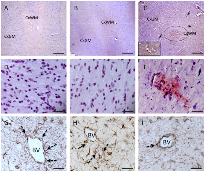
Figure 5. Photomicrograph showing changes to the cerebrovasculature following UCO.
Panels A–C show albumin staining in the cortical gray (CxGM) and subcortical white matter (CxWM) of a full term (A), a preterm (B) and a UCO lambs (C). Albumin extravasation (brown staining) consistent with blood brain barrier permeability disruption was observed throughout the brain in UCO lambs (C). Note the positive albumin staining around a blood vessels (circle), as well as in cells (arrows). The insert in C is a high power photomicrograph showing the albumin staining surrounding a blood vessel (BV), as well as albumin positive cells, We observed moderate levels of albumin extravasation in some pre tem control lambs (B), while no albumin staining was noted in brains of full term control lambs. Panels D, E and F show Mallory trichrome staining in the periventricular white matter of a full term (D), preterm (E) and UCO lamb (F). Microbleeds were seen in UCO brains shown by degradation products of hemorrhage staining a muddy brown. No microbleeds were detected in full term or preterm control brains. G–I show GFAP immunohistochemistry and show normal perivascular astrocytes in the periventricular white matter of a full term (G) and preterm lamb (H); while blood vessels in UCO lamb were often seen to be devoid of astrocytic contact (I). BV = blood vessel. Scale bars: A–C = 100 µm; D–I = 20 µm.

