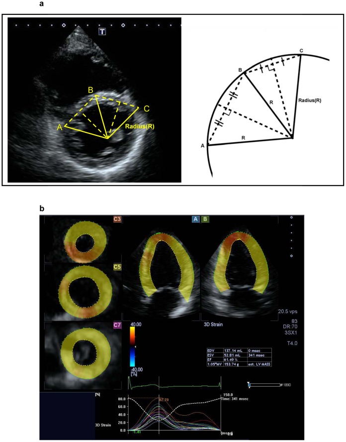
Figure 1. Two-dimensional echocardiographic assessment of ventricular septal configuration and 3D speckle tracking analysis.
(a) The radius was identification from the intersection of perpendicular bisectors between the anterior (C) and middle (B) and between the posterior (A) and middle (B) points in the ventricular septum. (b) The 5-plane view used for 3D speckle tracking analysis: A plane (apical 4-chamber view), B plane (2-chamber view), and 3 C planes (short-axis views near the apex, at mid-level, and at the base of the left ventricle).

