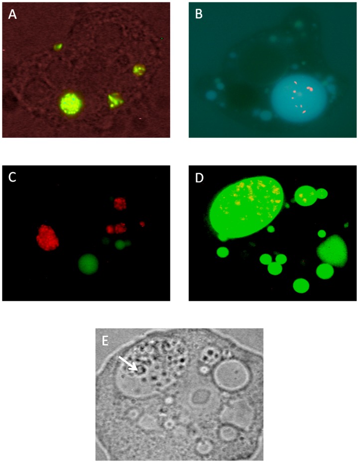
Figure 4. Viable and heat killed C. jejuni are taken up into different types of A. polyphaga vacuoles.
(A) Live/Dead stained viable (green) C. jejuni, confined to tight vacuoles. (B) Live/Dead stained heat killed (red) C. jejuni residing in giant spacious vacuoles. (C) Vaculoes with CTC stained viable (red) C. jejuni do not co-localize with Alexa fluor-488 labeled dextran filled vacuoles (green). (D) Vaculoes with CTC stained heat killed (red) C. jejuni have taken up Alexa fluor-488 labeled dextran (green). (E) In contrast to non digestive vacuoles, giant digestive vacuoles contained smaller vesicles (arrow). Picture D and E are from the same amoeba. ZEISS Axioskop (Germany)×63, FI 450–490 (FT 510, LP 520) and a Nikon camera COOLPIX 995 was used for fluorescence images A, C, D and bright field image E. OLYMPUS BX 50 (Japan) ×100 and an OLYMPUS camera DP 50 was used for fluorescence image B.

