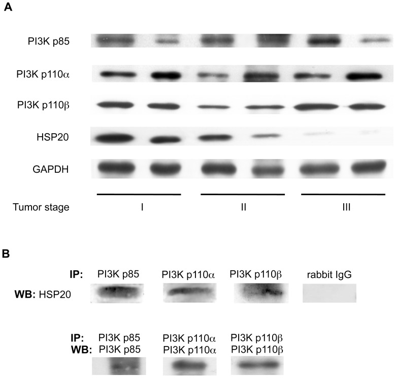
Figure 6. HSP20 interacts with PI3K in human HCC tissues.
(A) The protein levels of HSP20 and PI3K p85, PI3K p110α and PI3K p110β subunits in stages I, II and III human HCC tissues were compared by a Western blot analysis. (B) The lysates from a stage II HCC tissue were immunoprecipitated (IP) with antibodies for PI3K p85, PI3K p110α PI3K p110β or normal rabbit IgG followed by Western blotting (WB) using HSP20 antibodies. Immunoprecipitation of the PI3K p85, PI3K p110α and PI3K p110β subunits in the stage II HCC tissue lysates was confirmed by WB using PI3K p85 antibodies, PI3K p110α antibodies and PI3K p110β antibodies, respectively.

