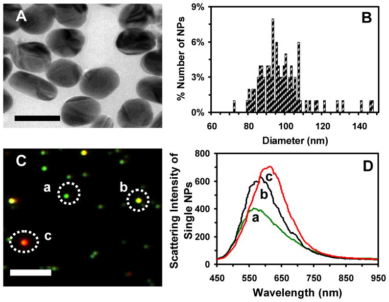
Figure 1.
Characterization of sizes, shapes and plasmonic optical properties of single Ag NPs suspended in egg water (24 pM) at 28.5°C over 120 h. (A) HRTEM images show polygon shaped NPs. (B) Histogram of the size distribution of single Ag NPs measured by HRTEM shows the average sizes of (97 ± 13) nm. The size of each NP is determined by averaging its length and width. (C) Dark-field optical image of single Ag NPs shows individual green, yellow and red/orange NPs. (D) LSPR spectra of single Ag NPs in (C) show λmax (FWHM): (a) 565 (146); (b) 585 (129); and (c) 615 (132) nm. Scale bars are 100 nm in (A) and 2 μm in (C). The scale bar in (C) shows the distances among individual NPs, but not their sizes due to the optical diffraction limit.

