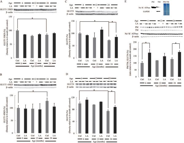
Fig. 2. Effect of lipoic acid on age-dependent changes in brain glucose transporters expression.
Equal amount of homogenate samples from brain cortices of Fischer 344 rats were loaded on the gel. Expression of (A) endothelial GLUT1 55 kDa; (B) glial GLUT1 45 kDa; (C) neuronal GLUT3; (D) neuronal GLUT4. (E) Lipoic acid induced the translocation of GLUT4 from the cytosol to plasma membrane. GLUT4 translocation was assessed by the relative expression of GLUT4 on the plasma membrane fraction and total membrane. Na+/K+ ATPase and β-actin were used as loading control for plasma membrane and total membrane, respectively. Top panel: the purity of plasma membrane fraction was determined by Na+/K+ ATPase and GAPDH. *p < 0.05, n ≥ 6.

