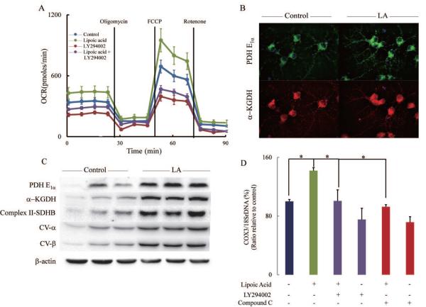
Fig. 4. Bioenergetics of primary cortical neurons.
(A) Cells were treated with control vehicle, R-(+)-lipoic acid (20 μM), LY294002 (50 μM), and R-(+) lipoic acid (20 μM) + LY294002 (50 μM) for 18 h. Oxygen consumption rate (OCR) was determined using Seahorse XF-24 as described in the Methods section. (B) Lipoic acid increased expression of PDH-E1α and α-KGDH. Cells were treated with vehicle or R-(+)-lipoic acid (20 μM) for 18 h before they were subjected to immuno-fluorescent staining. (C) Lipoic acid increased expression of Complex II-SDHB, CV-α, CV-β, PDH-E1α and α-KGDH. After treated with vehicle or R-(+)-lipoic acid (20 μM) for 18 h, cells were harvested and lysed in Mammalian Protein Extraction Reagent (M-PER). (D) Lipoic acid increased mitochondrial density and was sensitive to PI3K (LY294002) and AMPK (compound C) inhibitors. *p < 0.05, n = 5 wells per group.

