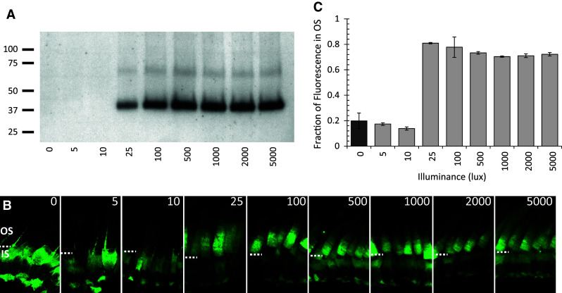
Fig. 9.

Phosphorylation of BBS5 correlates with the same light intensity that initiates translocation of Arr1 to the outer segments. a One eye from each Arr1–GFP tadpole was incubated with 32P-γATP and was exposed to increasing intensities of light (0–5,000 lux) for 15 min, homogenized, separated by SDS-PAGE, and autoradiographed; molecular mass standards (in kDa) are indicated to the left. b The contralateral eye was exposed to the same lighting and then fixed for imaging of GFP fluorescence in the photoreceptors by confocal microscopy. The dashed white line indicates the boundary between the outer segments (OS) and inner segments (IS). c Quantification of Arr1 translocation to the outer segments; the fraction of GFP fluorescence in the OS was measured and averaged from a minimum of 15 photoreceptors in two separate sections from each of two eyes
