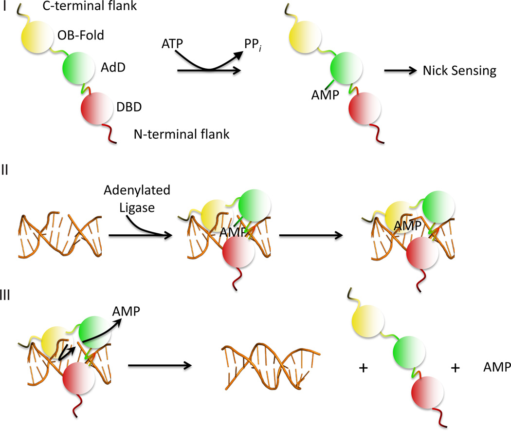
Figure 1.
Steps in the DNA ligation reaction. (I) The catalytic region of the DNA ligase consisting of the DNA binding domain (DBD, red), adenylation domain (AdD, green) and oligonucleotide/oligosaccharide binding-fold (OB-Fold, yellow), interacts with ATP to adenylate an active site lysine within the adenylation domain (AdD, green), releasing pyrophosphate; (II) When the adenylated ligase recognizes and binds to a DNA nick, it undergoes a conformational change such that the DBD, AdD and OB-fold encircle the nick. Within this compact structure, the AMP moiety is transferred from the ligase polypeptide to the 5' phosphate of the nick; (III) The non-adenylated ligase polypeptide utilizes the 3' hydroxyl terminus of the nick as a nucleophile to attack the 5' DNA-adenylate, resulting in phosphodiester bond formation and the release of the ligase polypeptide and AMP

