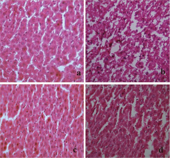
Figure 5. Administration of ACP/silymarin to the groups intoxicated with APAP.

(a): Section of liver of normal control rats showing hepatic cells with well defined nuclei and cytoplasm (350×). (b): Section of APAP treated rat liver showing marked necrosis, extensive vacuolation, broad infiltration of lymphocytes and Kupffer cells, loss of cell boundaries and disappearance of nuclei (350×). (c): Section of APAP+ACP (150 mg/kg) treated rat liver showing marked improvement over APAP control group (350×). (d): Section of APAP+silymarin (100 mg/kg) treated rat liver showing normalcy of hepatic cells (350×).
