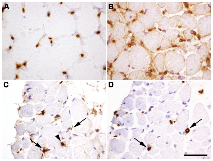
Figure 5. Characterization of inflammation found in UCMD muscle.
HLA immunostaining in healthy muscle sections located in the endothelial cells of capillaries (A). Sarcolemmal and cytoplasmatic HLA staining on UCMD muscle sections including strong staining of mononucleated cells (B). Immunohistochemistry for CD68 demonstrate macrophage infiltration (C). Immunohistochemistry for CD206 identified M2-type macrophages (D). Arrows point to mononucleated cells CD68+ and CD206+ whereas arrowheads point to only CD68+ cells. Scale bar: 50 µm.

