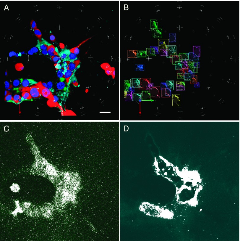
Figure 1.
Active cysteine cathepsins and degradation fragments of DQ-collagen IV could be detected in breast carcinoma cells grown in 3D rBM overlay cultures. (A) 3D reconstruction of optical sections taken through the entire volume of an MDA-MB-231 3D culture: with red representing RFP in the cytoplasm; blue representing Hoechst 33342-stained nuclei; green representing degradation products of DQ-collagen IV; and cyan representing GB123 bound to active cysteine cathepsins. (B) Illustration of spatial separation in 3D of nuclei for enumeration. (C and D) In single optical sections through the equatorial plane, fluorescence was evenly increased to visually differentiate with mask (white), illustrating measured DQ-collagen IV degradation fragments and ABPs bound to cysteine cathepsins, respectively. Bar, 22.6 µm.

