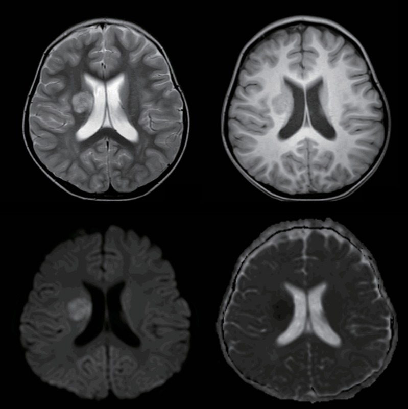
Fig. 1.
Magnetic resonance imaging (MRI) was performed on a 3T MRI scanner (Achieva 3T; Philips Medical System). MRI images showed a round lesion involving the right basal ganglia, which was hyperintense on T2-weighted images (top left image) and hypointense on T1-weighted images (top right image), with diffusion restriction (bottom left image) and low apparent deficient coefficient values (bottom right image), indicating acute infarction in the area of the right lenticulostriate arteries.

