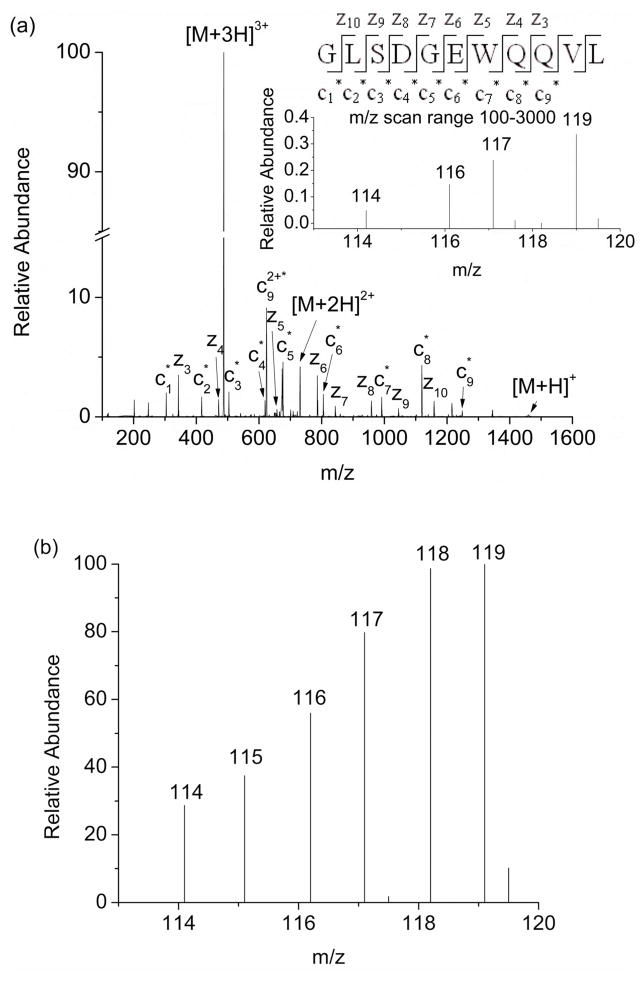
Figure 4.
(a) ETD mass spectrum of the [M+3H+TMT6]3+ ion of the peptide fragment GLSDGEWQQVL. A series of modified c1 to c9 ions and a series of unmodified z3 to z10 ions confirm that the N-terminus is the site of modification. The product ions with an asterisk are the product ions that contain the TMT modification. The inset spectrum is an enlarged view of the reporter ion region, showing that poor ion statistics leads to insufficient information to construct a reliable dose-response plot. (b) ETD mass spectrum of the [M+3H+TMT6]3+ ion of the peptide fragment GLSDGEWQQVL, acquired using a shorter scan range (from m/z 100 to 150).

