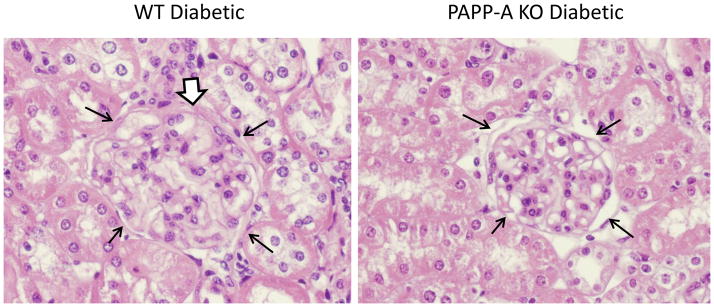
Figure 2. Renal histology of diabetic WT and PAPP-A KO mice.

Representative H & E staining of kidney sections. Thin black arrows delineate a glomerulus. White-filled arrow indicates thickened Bowman’s capsule.

Representative H & E staining of kidney sections. Thin black arrows delineate a glomerulus. White-filled arrow indicates thickened Bowman’s capsule.