Figure 3.

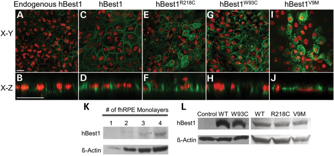
Localization of endogenous hBest1, overexpressed hBest1, and overexpressed hBest1 mutants (R218C, W93C, and V9M) in fhRPE cells. Representative X–Y and X–Z scans of hBest1 (green) localization are shown for endogenous hBest1 (A and B) or following adenovirus-mediated gene transfer, for overexpressed hBest1 (C and D), hBest1R218C(E and F), hBest1W93C(G and H), or hBest1V9M(I and J) in polarized monolayers of fhRPE cells. Nuclei (red) were used as a positional marker. Like endogenous hBest1, overexpressed hBest1, hBest1R218C and hBest1W93C localized to the basolateral plasma membrane, while hBest1V9M remained intracellular. Scale bars: 20 µm. (K) Western blotting of fhRPE monolayers revealed endogenous expression of hBest1 with β-actin as a loading control. (L) Western blotting of hBest1 and β-actin in fhRPE cells (control) or fhRPE cells overexpressing hBest1 or hBest1 mutants following adenovirus-mediated gene transfer demonstrates level of overexpression of hBest1 in infected cells.
