Figure 5.

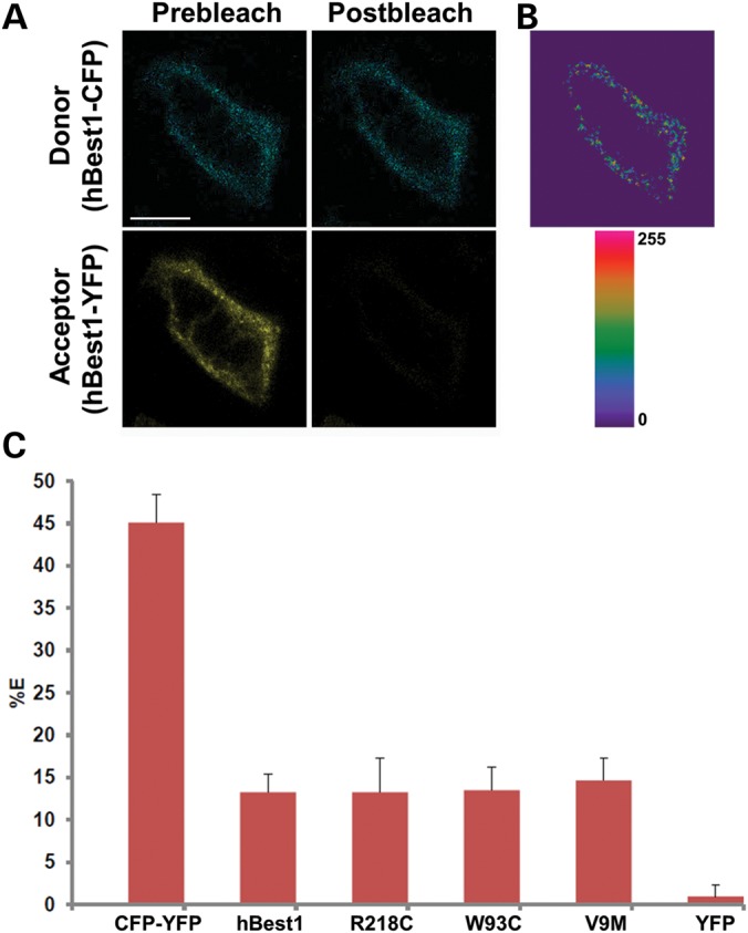
Live-cell, confocal FRET acceptor photobleaching of hBest1-CFP and hBest1-YFP or mutant (R218C, W93C and V9M) hBest1-YFP in MDCK II cells. (A) Representative X–Y scan of hBest1-CFP (blue, donor) and hBest1-YFP (yellow, acceptor) co-expressed in confluent MDCK II cells using adenovirus-mediated gene transfer. Live-cell acceptor photobleaching was performed by bleaching the acceptor, generating the resultant image in (B), which highlights regions in the plasma membrane where donor intensity increased. (C) FRET efficiencies (%E's) were determined for hBest1-CFP paired with hBest1-YFP (n = 23) or hBest1V9M-YFP (n = 23) via adenovirus-mediated gene transfer or hBest1W93C-YFP (n = 21) or hBest1R218C-YFP (n = 24) hBest1 via transfection. MDCK II cells were transfected with a CFP–YFP fusion protein (n = 26) as a positive control and hBest1-CFP and YFP (n = 25) as a negative control. Both hBest1 and mutant hBest1 had %E's significantly different (P < 0.001) than the negative and positive controls. Scale bar: 10 µm. Error bars indicate ± SD.
