Figure 7.
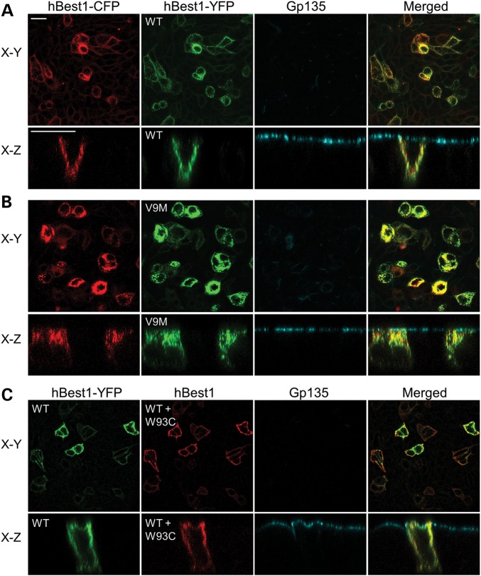
Effect of hBest1 on localization of hBest1V9M, or hBest1W93C in MDCK II cells. Polarized monolayers of MDCK II cells were made to co-express hBest1-CFP and hBest1-YFP, hBest1V9M-YFP, or hBest1W93C via adenovirus-mediated gene transfer. Gp135 (cyan) was used as an apical protein marker for positional referencing. Representative X–Y and X–Z scans are shown for each co-localization experiment. (A) Both hBest1-CFP (red) and hBest1-YFP (green) co-localized to the basolateral plasma membrane. (B) Both hBest1-CFP (red) and hBest1V9M-YFP (green) co-localized in intracellular compartments. (C) hBest1-YFP (green) was co-expressed with untagged hBest1W93C and cells were stained for hBest1 (red). hBest1 staining was in the basolateral plasma membrane, indicating that the presence of WT hBest1 rescued the mislocalization of hBest1W93C. Scale bars: 20 µm.

