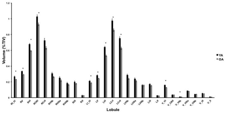
Figure 3.
Age differences in lobular volume (YA: black bars; OA: gray bars). There are significant main effects of age (F(1,51)=20.17, p<.001) indicating that older adults have smaller cerebellar gray matter volume. There is also a main effect of lobule (F(4.26,217.14)=1985.66, p<.001), along with a significant age by lobule interaction (F(4.26,217.14)=9.07, p<.001). *Indicates significant age differences in lobular volume as assessed using follow-up pairwise t-tests, corrected for multiple comparisons (p<.002). Error bars represent the standard error of the mean. Lobules are labeled using roman numerals. L: left hemisphere; R: right Hemisphere; V: vermis; CRI: Crus I; CRII: Crus II.

