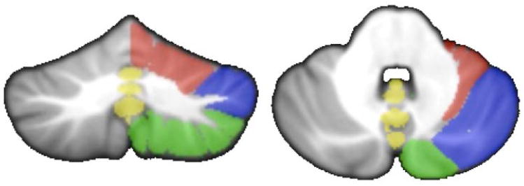
Figure 5.
PCA components in the young adults were used to define sub-regions of the cerebellum for additional analysis. Overlays are presented on the right hemisphere only on both a coronal (left) and axial (right slice), to allow for comparison to the left hemisphere, though analysis was conducted on both hemispheres. Lobules not included in a component (left and right lobule X and right Crus II) were included in the posterior grouping given their anatomical location. Red: anterior cerebellum, corresponding to component two; Green: posterior cerebellar grouping corresponding to component one; Blue: Crus I, corresponding to component four; Yellow: cerebellar vermis corresponding to component three.

