
Figure 6.
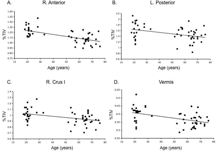
Correlations with age and regional cerebellar volume (% TIV). A) right anterior cerebellum (r=−.64), B) left posterior cerebellum (r=−.38), C) right Crus I (r=−.41), and D) the vermis (r=−.47). Additionally, there were significant negative correlations with the left anterior cerebellum (r=−.64) and left Crus (r=−.48), though just the right hemisphere is presented here for the sake of parsimony. These linear relationships indicate differences in cerebellar volume across adulthood.