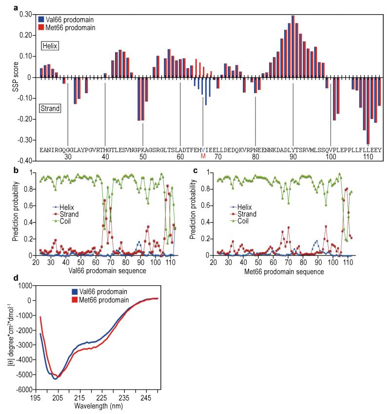
Figure 4.
Val66 and Met66 prodomains differ in transient secondary structure.(a) Secondary structure propensity (SSP) score obtained using the backbone 1H, 15N, 13C chemical shifts from the BDNF prodomain Val66 (blue bars) and Met66 (red bars). The SSP score identified regions of transient structure formation (positive values = α-helix, negative values = β-sheet). (b)(c) Graphs illustrating secondary structure prediction by TALOS+ analysis using the heteronuclear backbone chemical shifts of Val66 (b) and Met66 (c) prodomains. α-helix in blue spheres, β-strand in red squares, and disorder in green triangles. TALOS+ analysis showed decreased β-sheet propensity in the Met66 prodomain compared to the Val66 prodomain, result that is consistent with the SSP score. (d) Ultraviolet circular dichroism (CD) spectra of 30 μM of the Val66 (blue) and Met66 (red) prodomains collected in10 mM NaH2PO4 and 50 mM NaCl pH 7.0 at 23°C. The negative peak around 200 nm revealed the natively unfolded conformation of both prodomains. However, the lower absorption at 222 nm for Met66 prodomain is consistent with increased tendency to helical conformation compared to the Val66 prodomain. Each spectrum is representative of 4 averaged scans and is normalized to the spectrum of buffer alone.

