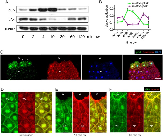
Fig. 2.
ERK and PI3K signalling are sequentially activated during embryonic wound healing. (A) Western blot analysis shows the sequential activation of pERK and pAkt in deep wounds, with α-tubulin as loading control. (B) Quantification of pERK and pAkt activation during embryonic wound healing. Data are means ± s.e.m. from three independent experiments and represent pERK and pAkt signal intensities normalized to α-tubulin controls. (C) Immunofluorescence staining of pERK, β-catenin (plasma membrane) and DAPI (nucleus) on a transected embryonic wound 10 minutes after wounding. w, wound; bl, blastocoel; ep, epithelium. (D–F) PIP3 localisation in unwounded epithelium (D), early phase wound (E) and late phase wound (F). Green, GFP-Grp1; red, mCherry-moesin; w, wound; ep, epithelium; pw, post wounding. Scale bars: 50 µm (C), 20 µm (D–F).

