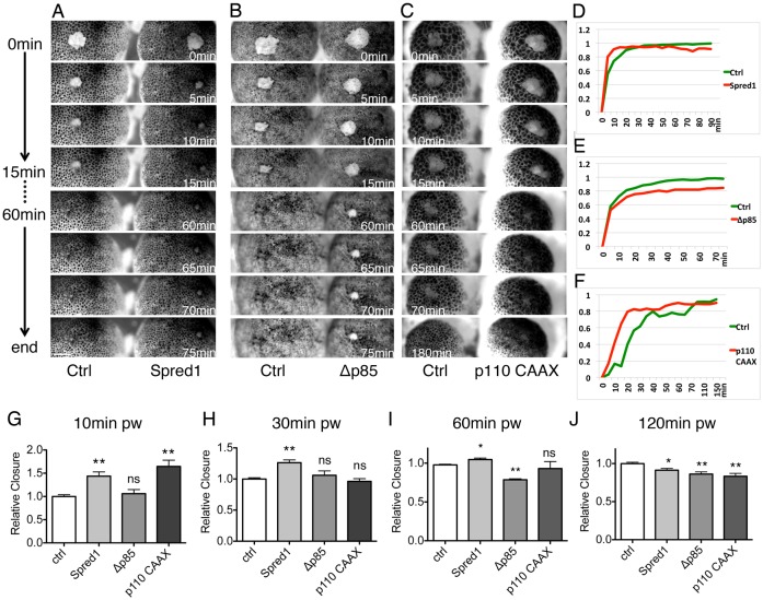
Fig. 3.
ERK and PI3K signalling regulate distinct phases of wound healing. (A–C) Time-lapse series of wound healing in control and in embryos injected with spred1 (A), Δp85 (B) and p110 caax (C). Each image shows time post wounding. Scale bar: 200 µm. (D–F) Healing curves of results in A–C. (G–J) Quantification of relative wound closure 10 minutes (G), 30 minutes (H), 60 minutes (I) and 120 minutes (J) post wounding. Mean wound closure percentage of control embryos was normalized to 1; other groups show closure relative to control. Results are shown as means ± s.e.m.; n = 3. Non-parametric Mann–Whitney test was used to test for significance between control and other groups. *P<0.05; **P<0.01; ***P<0.001; ns, not significant.

