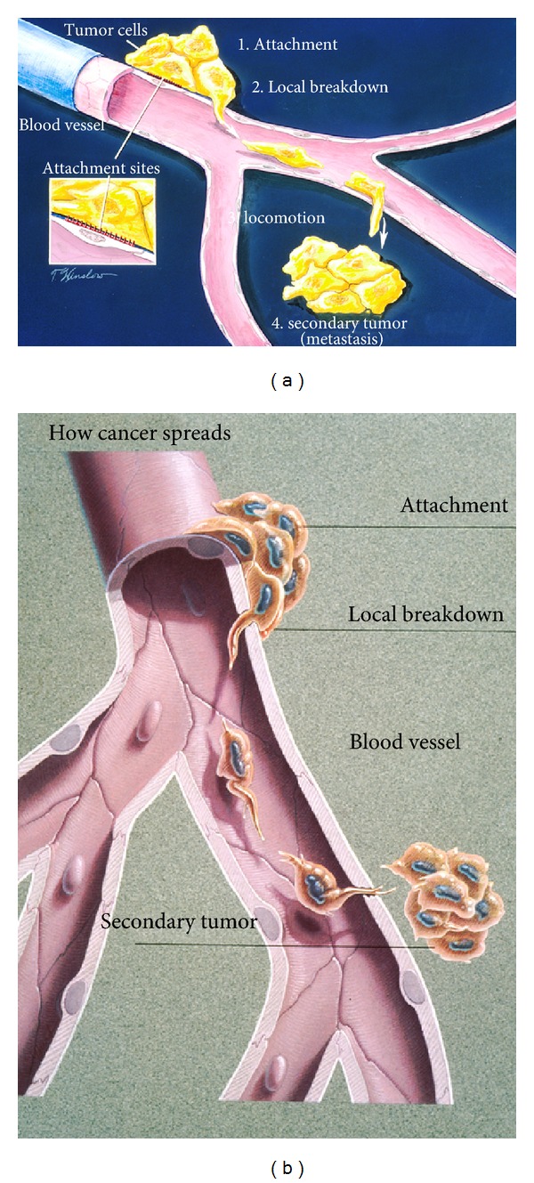
Figure 10.

(a) Schematic drawing for the stages of metastasis (1) attachment (2) local breakdown with lamellipodia penetration into a blood vessel (3) locomotion with lamellipodia to exit from blood vessel (4) secondary tumor. (b) Once metastatic cells are attached to the vessel wall basement membrane (a physical barrier that separates tissue components), cancer can break through with stiff lamellipodia on the leading edge and the help of protease enzymes. Cancer cells then move through the blood stream enabling them to spread to other parts of the body. A secondary tumor may subsequently form at another site in the body. (With permission from the National Institutes of Health/Department of Health and Human Services).
