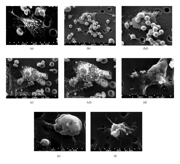
Figure 12.

Six-step sequence showing the death of a cancer cell. (a) One cancer cell is migrating through a hole of a matrix-coated membrane from the top to the bottom, simulating natural migration of an invading cancer cell between, and sometimes through, the vascular endothelium. Notice the spikes or pseudopodia that are characteristic of an invading cancer cell. A film coat containing red blood cells, lymphocytes, and macrophages is added to the bottom of the membrane. (b) A group of macrophages identify the cancer cell as foreign matter and start to stick to the cancer cell, which still has its spikes. (b2) enlarged for relative cancer cell size comparisons for approximate equal magnifications between Figures (a), (b2), (c2), (d), (e) inset lower left, and (f). (c) Macrophages begin to fuse with, and inject toxins into, the cancer cell. The cancer cell starts rounding up and loses its spikes. (c2) enlarged for cancer cell size comparisons. (d) As the macrophage cells become smooth, the cancer cell appears lumpy in the last stage before it dies. (e) Lumps covering the cancer cell surface are actually the macrophages fused within the cancer cell with inset at lower left for cancer cell size comparisons showing a great reduction in size. (f) The cancer cell then loses its morphology, shrinks up more, and dies. (With permission from the National Institutes of Health/Department of Health and Human Services).
