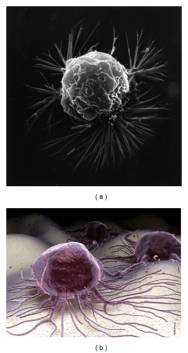
Figure 9.

(a) SEM of a breast cancer cell that clearly shows both the soft petals of membrane ruffling and much longer lamellipodia spiking extensions. (b) A scanning electron microscopic 3D-enhanced NIH image of cancer cells and lamellipodia spike processes on a cellular tissue surface (with permission from the National Institutes of Health/Department of Health and Human Services).
