
Figure 6.
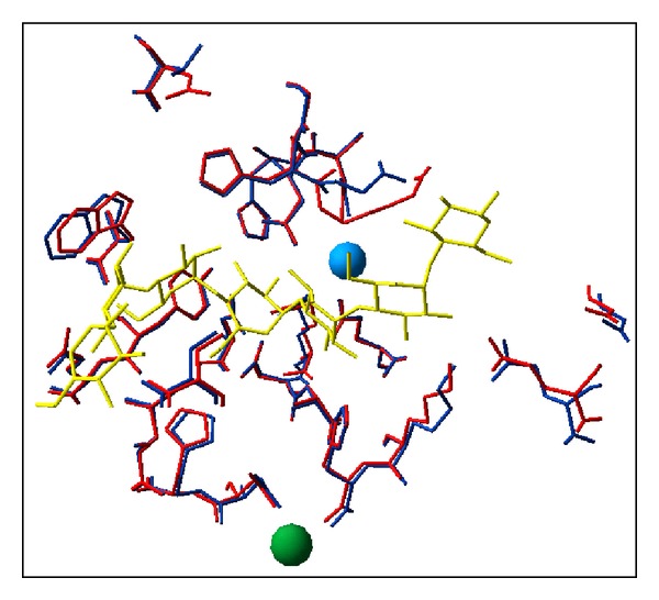
Structure of the active site. Superimposition of the active site residues in psychrophilic (blue) and mesophilic α-amylases (red). The chloride and calcium ions are shown as blue and green spheres, respectively. The 24 residues performing direct or water-mediated interactions with a substrate analog (yellow) are identical and superimpose almost perfectly within the resolution of the structures, demonstrating a structural identity in these psychrophilic and mesophilic enzymes [59].