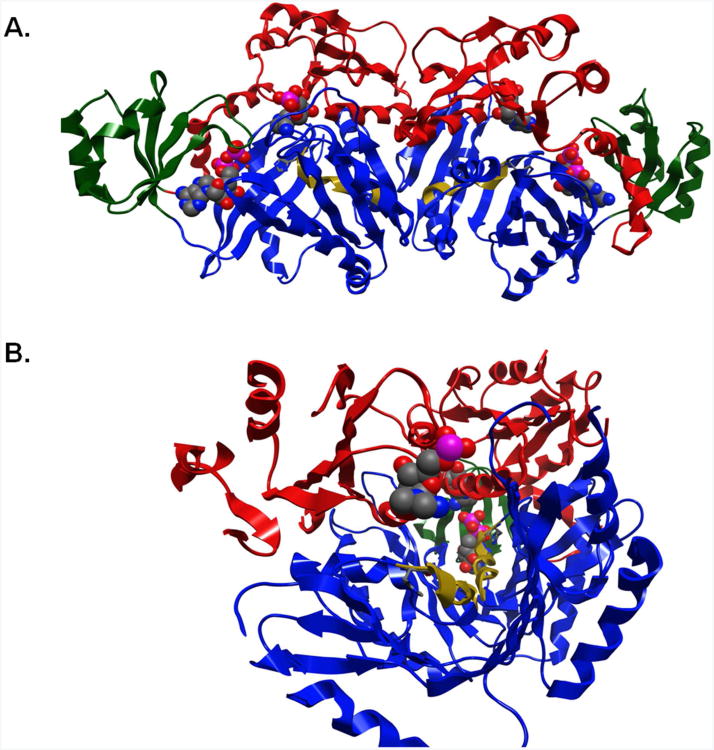
Figure 5. Structure of N5-CAIR synthetase dimer.
A. The A, B, and C domains of each subunit are shown in red, green and blue respectively. AIR and ATP are shown in spacefilling representation, while H273 is shown in the stick representation. The H273-E-281 helix is shown in gold. B. The view down the H273-E281 helix. Notice that the two helices (gold) are pointing at the other active site. The AIR of one subunit is shown in the front, while the second is shown in the center behind the second helix. This picture was created by rotating to the B-domain and Z-clipping the domain to gain access to the AIR binding site.

