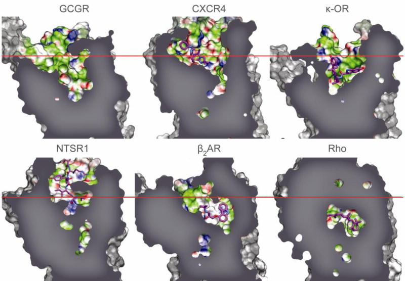
Figure 2. Comparison of ligand binding pocket of GCGR with class A GPCRs.
The binding cavity of GCGR compared with the binding cavities of human chemokine receptor CXCR4 (PDB 3ODU), human κ-opioid receptor (κ-OR) (PDB 4DJH), rat neurotensin receptor (NTSR1) (PDB 4GRV), human β2 adrenergic receptor (β2AR) (PDB 2RH1), and bovine rhodopsin (Rho) (PDB 1U19) for comparison purposes (Supplementary Table 4). The approximate position of the EC membrane boundary is shown as a red line, and bound ligands as magenta carbon atoms.

