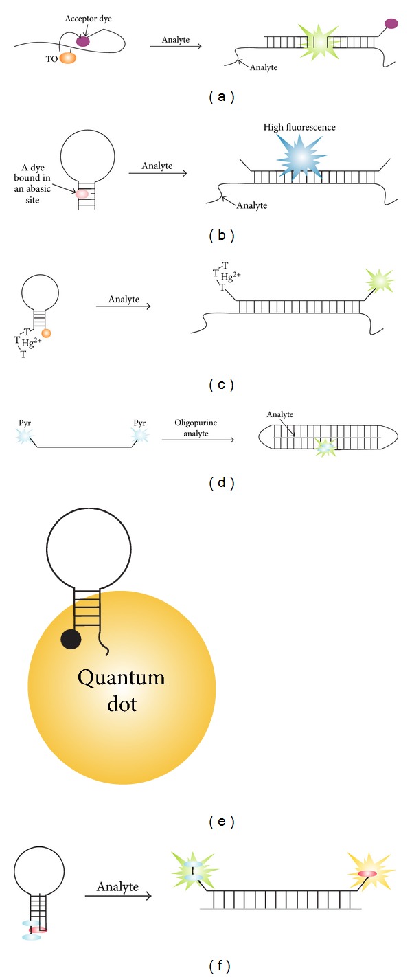
Figure 12.

Recent variations of MB probes. (a) Dual fluorophore PNA FIT probes [116]. The flexibility of a single-stranded PNA chain enables the contact of thiazole orange dye (TO) with the acceptor dye (fluorescent or dark quencher), which results in efficient quenching of TO fluorescence. The fluorescence increase upon target binding is achieved due to (i) separation of the fluorescent donor from the fluorescent acceptor and (ii) increase in fluorescence as a result of TO stacking interactions with newly formed base pairs. (b) Label-free MB probe (APMB) [117]. When noncovalently bound to an abasic site within the stem structure of the probe, a dye produces low fluorescent signal. Hybridization to a complementary target releases the dye free in solution, thus enabling high fluorescence. (c) Thymidine-terminated MB probe [118]. The fluorescence of FAM group in the closed conformation of the probe was quenched due to the binding of Hg2+ ions by three consecutive thymidine residues on the opposite terminus of the probe. (d) Triplex-forming DNA probes [116]. (e) Quantum dot-(QD-) based MB probe [117]. (f) Two-color MB probe [119]. Cyan ovals are two pyrene residues, which interact with perylenediimide residue (red). Hybridization of an analyte separates pyrene dimer form perylenediimide, thus increasing the fluorescence of both dyes.
