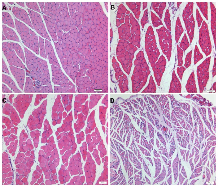
Figure 3. Photomicrographs of transverse sections of the gastrocnemius muscle following denervation and subsequent reinnervation.
The muscle sections of (A) immediate repair, (B) chronic denervation with sensory protection and (C) chronic denervation with mixed-nerve protection showed wide areas of larger muscle fibers among small groups of atrophied fibers. (D) Unprotected muscle sections showed extensive atrophy. Magnification×100. Scale bar= 100µm.

