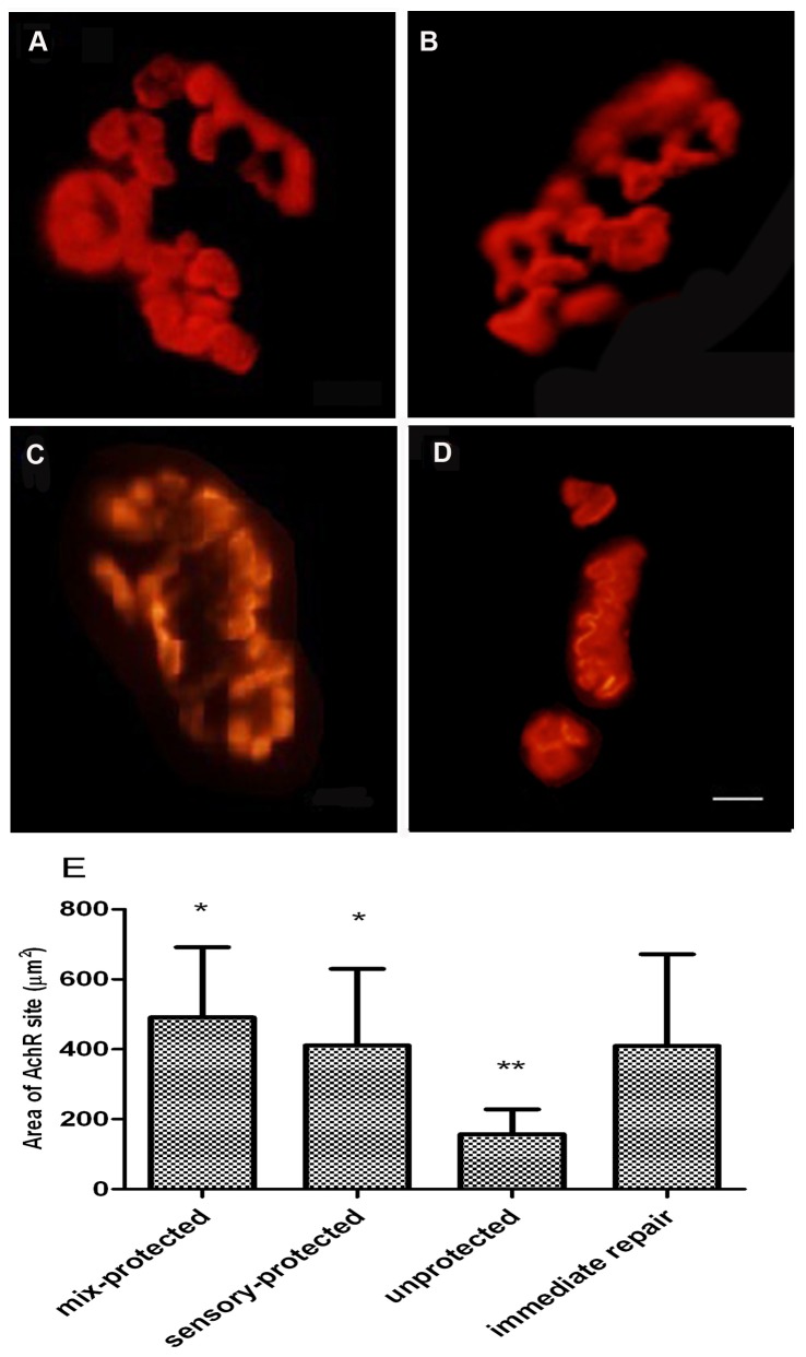
Figure 4. Postsynaptic AChR plaques at motor endplates in gastrocnemius muscle, as visualized by α-bungarotoxin binding.
(A) AChR plaques in the immediate repair group had well-defined contours. The appearance of AChR plaques in (B) chronic denervation with sensory protection and (C) chronic denervation with mixed-nerve protection displayed thick fringes and a few small round cupulae with distinct contours. The appearance of AChR plaques in (D) the unprotected group appeared as delineated, small, flat and slender. Magnification×400. Scale bar= 5µm. (E) Graphs showed that the area of AChR sites in the unprotected group was significantly smaller than in the other groups. (*p < 0.05 compared with unprotected control; **p < 0.05 compared with immediate-repair control).

