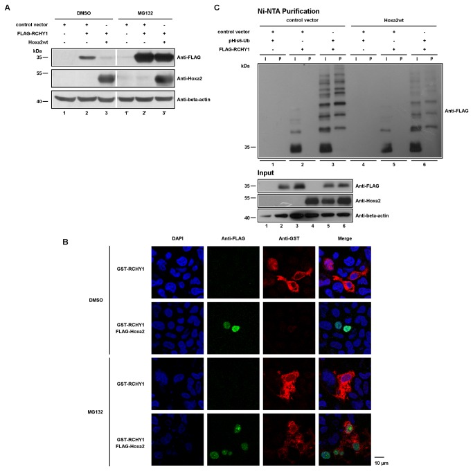
Figure 2. Hoxa2 induces proteasome-dependent ubiquitin-independent RCHY1 degradation and colocalizes with RCHY1 in the nucleus.
(A) HEK293T cells were co-transfected with expression vectors for Hoxa2 and FLAG-tagged RCHY1, and treated with MG132 proteasome inhibitor, or DMSO. Cell lysates were loaded on SDS-PAGE for proteins separation and western blotting (beta-actin used as a protein load control). (B) FLAG-Hoxa2 and GST-RCHY1 colocalize in the nucleus. HEK293T cells were co-transfected with FLAG-tagged Hoxa2 and GST-tagged RCHY1, treated with MG132 proteasome inhibitor, or DMSO, and subjected to immunocytochemistry with anti-FLAG M2 antibody (green) and anti-GST antibody (red). Nuclei were stained with DAPI (blue). (C) Ubiquitination assays for FLAG-tagged RCHY1. HEK293T cells were co-transfected with indicated expression vectors and treated with MG132. Cells were then lysed and 6His-ubiquitin conjugated proteins were purified using Ni-NTA beads. Purified proteins and cell lysates were analysed by western blotting using M2 antibody to detect ubiquitinated forms of FLAG-tagged RCHY1. Lysate samples were loaded on a SDS-PAGE to verify protein levels prior to purification (Input; beta-actin was used as a protein load control). Lane numbering under the gels identifies cell samples. I: input sample; P: Ni-NTA purified sample.

