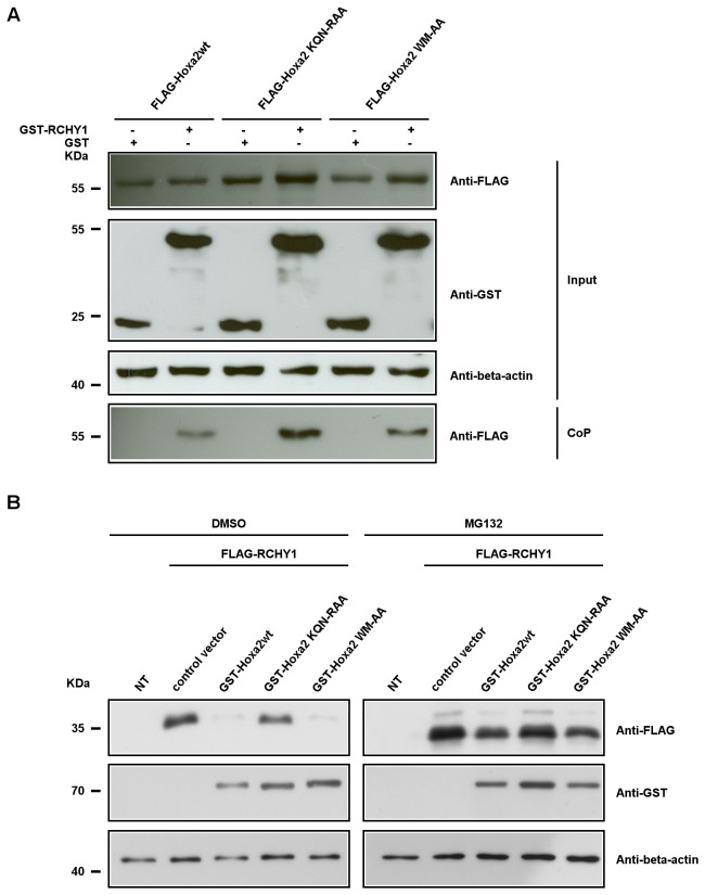
Figure 3. The integrity of the Hoxa2 homeodomain is required for the Hoxa2-induced RCHY1 decay.
(A) Co-precipitation assays involving mutant forms of Hoxa2. HEK293T cells were cotransfected with expression vectors for FLAG-tagged Hoxa2 (FLAG-Hoxa2wt), FLAG-tagged Hoxa2KQN-RAA (FLAG-Hoxa2KQN-RAA), FLAG-tagged Hoxa2WM-AA (FLAG-Hoxa2WM-AA), GST-tagged RCHY1 and GST proteins, and treated with the proteasome inhibitor MG132. Forty-eight hours after transfection, cell lysates were subjected to western blotting (input) and protein interactions were verified by co-precipitation on glutathione beads directed toward the GST tag. Eluted proteins were analysed by western blotting using the M2 anti-FLAG antibody (CoP). (B) Amino acid substitutions in the Hoxa2 homeodomain abolish the Hoxa2-mediated degradation of RCHY1. HEK293T cells were transfected with expression vectors for FLAG-tagged RCHY1 (FLAG-RCHY1) and GST-tagged Hoxa2 (GST-Hoxa2wt, GST-Hoxa2KQN-RAA, GST-Hoxa2WM-AA) proteins. Cells were then treated for proteasome inhibition (MG132) and compared to untreated controls (DMSO) for the decay of FLAG-RCHY1 revealed by western blot detection. Detection of beta-actin was used as a protein load control.

