
Figure 3.
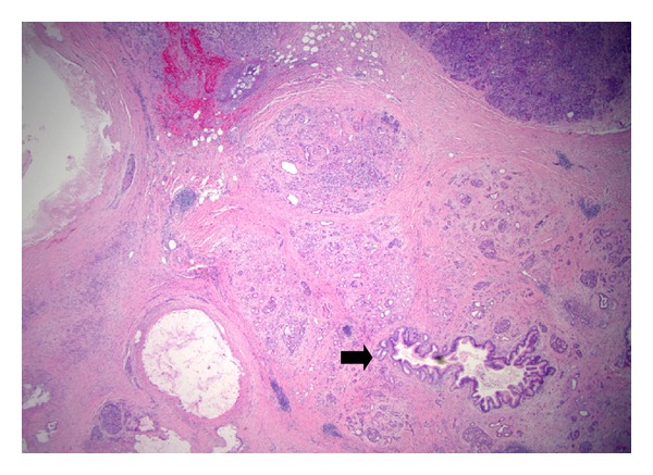
Atrophy of parenchyma associated with IPMN. The acinar parenchyma in the vicinity of an IPMN (not shown) exhibits loss of lobular acini, with a central duct and islets of Langerhans remained. Note the small duct (arrow) is secondarily involved by changes of IPMN, with microscopic papillary overgrowth of the epithelium.