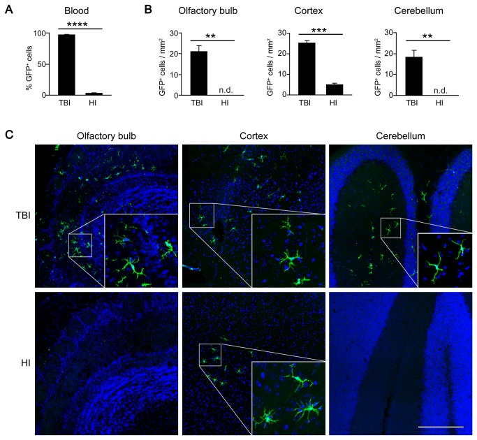
Figure 2. Delayed engraftment of BMDCs after HI.
A) Flow cytometric analysis of GFP expression in peripheral blood leukocytes of HI and TBI chimeras. The level of chimerism was significantly lower in HI compared with TBI animals at 16 weeks after transplantation. Data are means + SEM from 3-5 animals per group. Statistical significance is indicated by the asterisk (****p<0.0001). B) Quantification of BMDC engraftment in the brains of HI and TBI chimeras. Data are expressed as GFP+ cells per area in three different brain regions (olfactory bulb, cortex and cerebellum) at 16 weeks after BMT. Data are means + SEM from 3-5 animals per group. n.d. = none detected. Statistical significance is indicated by asterisks (**p<0.01; ***p<0.001). C) Representative laser confocal microscopic images of ramified donor-derived GFP+ cells in the brains of HI and TBI animals at 16 weeks after BMT. Note that GFP+ cells were restricted to the cortex in the HI group, but distributed throughout the grey and white matter in TBI animals.

