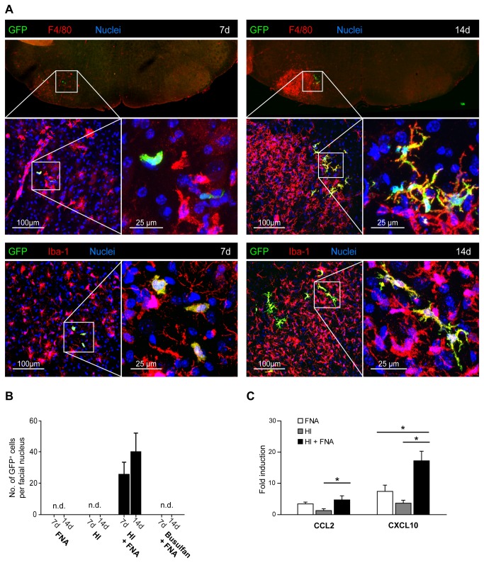
Figure 4. Selective engraftment of donor-derived myeloid cells in the lesioned facial nucleus after HI.
A) Identification of GFP+ myeloid cells in the lesioned facial nucleus of HI chimeras at 7 and 14 days after BMT. Note the increase in F4/80 immunoreactivity at day 14 compared with day 7, indicating increased inflammation. The contralateral unlesioned facial nucleus is devoid of donor-derived GFP+ cells and shows minimal F4/80 immunoreactivity. Laser confocal microscopic images of areas of interest (white squares) are shown at increasing magnifications (scale bars: 100 µm – 25 µm). Seven days after BMT, amoeboid GFP+F4/80+ and GFP+Iba-1+ cells were detected in the lesioned facial nucleus. At 14 days after BMT, GFP+F4/80+ and GFP+Iba-1+ cells in the lesioned facial nucleus were highly ramified. All donor-derived GFP+ cells expressed the macrophage markers, F4/80 and Iba-1. Nuclei were counterstained with DAPI. B) Quantification of myeloid cell engraftment in the lesioned facial nucleus at 7 and 14 days after BMT in FNA, HI, HI + FNA and busulfan + FNA animals. Note that donor-derived GFP+ cells were only detected in HI + FNA mice. Data are means + SEM from 3-5 animals per group. n.d. = none detected. C) Quantitative real-time PCR of CXCL10 and CCL2 mRNA expression in the facial nucleus of animals with FNA (white bars), HI (grey bars) and HI + FNA (black bars) at 14 days after BMT. The mRNA expression levels were normalized to GAPDH mRNA and compared to naïve mice (fold induction). Increased chemokine mRNA levels were observed in the facial nucleus of HI + FNA mice compared to HI animals. Data are means + SEM from 3-5 animals per group. Statistical significance is indicated by asterisks (*p<0.05).

